Thursday, 28 April 2016

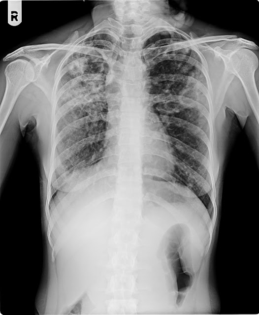
28/04/16 DHARMESHBHAI THAKOR M/34Y CXR

28/04/16 DHARMESHBHAI THAKOR M/34Y CXR
H/O ACUTE OEDEMATOUS PANCREATITIS..PT IS NBM.AND 170ML/HOUR FLUID IS GOING ON.BP -150/110 MM OH HG.DIABETES DETECTED FIRST TIME
NAME   :  DHARMESHBHAI  THAKOR  
AGE      :   34   Years.
REF BY:     DR.
 DATE   :   28/04/2016


X-RAY CHEST PA VIEW.


Inhomogenous opacity seen in LT lower zone possibility of consolidation.

Both apices, cardiophrenic, costophrenic angles and domes of  the diaphragm are normal.

The cardiac size is within normal limit.

Trachea is central, no mediastinal shift is seen and the  mediastinal outlines do not show any abnormality.

Bony thorax and soft tissues of the chest wall are normal.


IMPRESSION : - Inhomogenous opacity seen in LT lower zone possibility
of consolidation.


Thanks for reference


DR.BHAVESH PATEL     DR.NIRAV DESAI    DR.DEEPAK SHARMA    DR.JIGNESH PATEL
                 MD, DMRE.           MB,DMRD                   MD(Radio diagnosis)                MB,DMRD


Friday, 22 April 2016

23/0416 RAMILABEN PRAJAPATI F/42Y L.S-AP/LAT


23/0416 RAMILABEN PRAJAPATI F/42Y L.S-AP/LAT


H/O BACK PAIN LAST 5 DAYS
NAME   :  RAMILABEN
AGE       :  42 Years
REF BY  : DR.  SELF
DATE     :  23.04.2016

X-RAY LUMBO-SACRAL SPINE AP & LATERAL VIEW


No obvious lytic or sclerotic lesion.

The  lumber  vertebral bodies shows early  degenerative spondylotic changes in the form of osteophyte formation .

Rest of  The intervertebral disc spaces are maintained with normal heights and
no subchondral bony changes.

No evidence of lysis or lysthesis seen.

The intervertebral joints do not show any abnormality.

Pre and paravertebral soft tissues do not show any abnormality.


Thanks for reference

DR.BHAVESH PATEL     DR.NIRAV DESAI    DR.DEEPAK SHARMA    DR.JIGNESH PATEL
                 MD, DMRE.           MB,DMRD                   MD(Radio diagnosis)                MB,DMRD


--------------------------------------------------------------------------------------------------------------------------

--------------

Thursday, 21 April 2016

21/04/16 DAHYABHAI CHAUHAN M/55Y CXR

21/04/16 DAHYABHAI CHAUHAN M/55Y CXR

H/O KHASHI LAST ONE MONTH
NAME   :    DAHYABHAI  CHAUHAN
AGE      :     55  Years.
REF BY:     DR.
 DATE   :    21/04/2016


X-RAY CHEST PA VIEW.


Both lung fields shows prominent bronchovascular markings suggest changes of Bronchitis.

No evidence of koch’s lesion or consolidation seen.

Both apices, cardiophrenic, costophrenic angles and domes of  the diaphragm are normal.

The cardiac size is within normal limit.

Trachea is central, no mediastinal shift is seen and the  mediastinal outlines do not show any abnormality.

Bony thorax and soft tissues of the chest wall are normal.


IMPRESSION : - Both lung fields shows prominent bronchovascular markings suggest changes of Bronchitis.


Thanks for reference


DR.BHAVESH PATEL     DR.NIRAV DESAI    DR.DEEPAK SHARMA    DR.JIGNESH PATEL
                 MD, DMRE.           MB,DMRD                   MD(Radio diagnosis)                MB,DMRD


--------------------------------------------------------------------------------------------------------------------------

Wednesday, 20 April 2016

21/04/16 DHRUMIL CHAVDA M/4.5Y CXR

21/04/16 DHRUMIL CHAVDA M/4.5Y CXR

H/O KHASHI.COUGH LAST 1.5 YRS
NAME            :  DHRUMIL  CHAVDA  
AGE                :   4.5   Years.
REF BY          :    DR.
 DATE            :   21/04/2016


X-RAY CHEST PA VIEW.


Both lung fields shows prominent bronchovascular markings suggest changes of Bronchitis.
 Minimal Haziness seen in LT perihilar region possibility of consolidation.

Both apices, cardiophrenic, costophrenic angles and domes of  the diaphragm are normal.

The cardiac size is within normal limit.

Trachea is central, no mediastinal shift is seen and the  mediastinal outlines do not show any abnormality.

Bony thorax and soft tissues of the chest wall are normal.


IMPRESSION : - Both lung fields shows prominent bronchovascular markings suggest changes of Bronchitis.
 Minimal Haziness seen in LT perihilar region possibility of consolidation.


Thanks for reference


DR.BHAVESH PATEL     DR.NIRAV DESAI    DR.DEEPAK SHARMA    DR.JIGNESH PATEL
                 MD, DMRE.           MB,DMRD                   MD(Radio diagnosis)                MB,DMRD


--------------------------------------------------------------------------------------------------------------------------

Monday, 18 April 2016

18/04/16 DIMPLEBEN GOSAI F/33Y C.SPI-AP/LAT


18/04/16 DIMPLEBEN GOSAI F/33Y C.SPI-AP/LAT
NAME   :   DIMPLEBEN  GOSAI
AGE       :   33  Years
REF BY :   DR.
DATE    :   18/04/2016

X-RAY CERVICAL SPINE AP & LATERAL VIEW

P/o fusion of body of C6-C7 vertebrae noted.

The bone density is normal.

No obvious lytic or sclerotic lesion.

The  cervical  vertebral bodies and their  appendages  (pedicles and spinous  process) are normal.

The intervertebral disc spaces are maintained with normal heights and
no subchondral bony changes.

rest of The intervertebral joints do not show any abnormality.

The cranio vertebral junction is normal.

The  spinal alignment is maintained (with normal  lordotic  curve and
alignment of the anterior/posterior spinal lines).

Pre and para vertebral soft tissues do not show any abnormality.

No evidence of bony cervical ribs seen on either side.


IMPRESSION:    P/o fusion of body of C6-C7 vertebrae noted.

Thanks for reference

DR.BHAVESH PATEL     DR.NIRAV DESAI    DR.DEEPAK SHARMA    DR.JIGNESH PATEL
                 MD, DMRE.           MB,DMRD                   MD(Radio diagnosis)                MB,DMRD


--------------------------------------------------------------------------------------------------------------------------

18/0416 NARESHBHAI PANDYA M/58Y CXR-PA RT.LATERAL


18/0416 NARESHBHAI PANDYA M/58Y CXR-PA RT.LATERAL
H/O MOUTH BLEDING LAST 10 DAYS

NAME   :   NARESHBHAI 
AGE      :    58  Years.
REF BY:     DR.
 DATE   :   18/04/2016


X-RAY CHEST PA VIEW.   RT/LATERAL


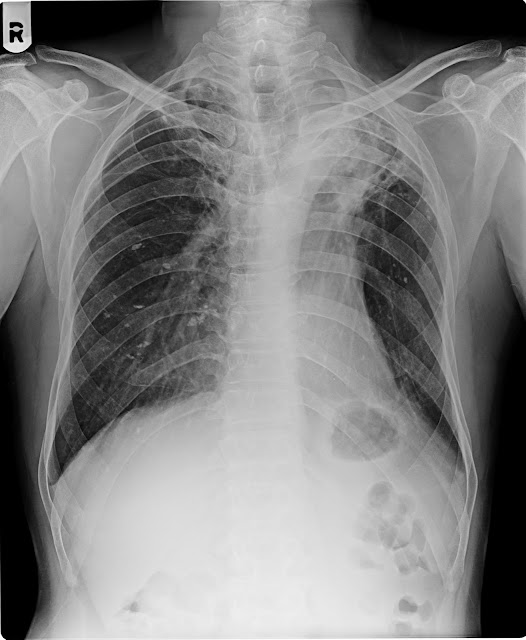
Soft tissue shadowing seen in left upper zone P/o Koch's consolidation.

Trachea shifted toward left side P/o under lying collapse.

Old calcified lesion seen in left upper & Rt mid lower zones.

Rest of both lung fields shows prominent bronchovascular markings.

 Both C P angle blunted P/o pleural thickening .

Bilateral apical pleural thiuckenong noted.

The cardiac size is within normal limit.

Bony thorax and soft tissues of the chest wall are normal.


Thanks for reference


DR.BHAVESH PATEL     DR.NIRAV DESAI    DR.DEEPAK SHARMA    DR.JIGNESH PATEL
                 MD, DMRE.           MB,DMRD                   MD(Radio diagnosis)                MB,DMRD


--------------------------------------------------------------------------------------------------------------------------

18/04/16 LAKHABHAI PARMAR M/55Y CXR

18/04/16 LAKHABHAI PARMAR M/55Y CXR
H/O MOUTH BLEDING LAST 15 DAYS

NAME            :   LAKHABHAI  PARMAR
AGE                :   55 Years.
REF BY          :   DR. 
 DATE            :   18/04/2016


  X-RAY CHEST PA VIEW.


Both lung fields shows prominent bronchovascular markings suggest changes of Bronchitis.

No evidence of koch’s lesion or consolidation seen.

Both apices, cardiophrenic, costophrenic angles and domes of  the diaphragm are normal.

The cardiac size is within normal limit.

Trachea is central, no mediastinal shift is seen and the  mediastinal outlines do not show any abnormality.

Bony thorax and soft tissues of the chest wall are normal.


IMPRESSION : - Both lung fields shows prominent bronchovascular markings suggest changes of Bronchitis.


Thanks for reference


DR.BHAVESH PATEL     DR.NIRAV DESAI    DR.DEEPAK SHARMA    DR.JIGNESH PATEL
                 MD, DMRE.           MB,DMRD                   MD(Radio diagnosis)                MB,DMRD


--------------------------------------------------------------------------------------------------------------------------

Sunday, 17 April 2016

18/04/16 DIMPLEBEN GOSAI F/33Y C.SPI-AP/LAT


18/04/16 DIMPLEBEN GOSAI F/33Y C.SPI-AP/LAT
H/O VERTIGO LAST 15 DAYS

-------------------------------------------------------------------------------------------------------------------------

Friday, 15 April 2016

16/04/16 MOHSIN ALI M/29Y CERVICAL SPINE-AP/LAT


16/04/16 MOHSIN ALI M/29Y CERVICAL SPINE-AP/LAT
H/O LT.SIDE HAND&LEG PAIN LAST ONE MONTH
NAME    :   MOHSIN ALI
AGE       :    29   YEARS
REF BY  : DR
DATE     : 16/04/2016
  
X-RAY OF CERVICAL SPINE (AP & LAT)       
--------------------------------------

Cervical spine appears normal.
Pedicles appear normal.
Disc spaces appear normal.
No evidence of  bony cervical ribs.
No evidence of wedging  is seen .
Pre and para  vertebral soft tissue shadow appears normal .

IMPRESSION ;
Normal x ray of cervical spine .


DR.BHAVESH PATEL     DR.NIRAV DESAI    DR.DEEPAK SHARMA    DR.JIGNESH PATEL
                 MD, DMRE.           MB,DMRD                   MD(Radio diagnosis)                MB,DMRD


-------------------------------------------------------------------------------------------------------------------------

15/04/16 REKHABEN TADVI F/30Y CXR-AP FOR RIBS

15/04/16 REKHABEN TADVI F/30Y CXR-AP FOR RIBS

H/O TRUMA LAST 2 DAYS LT.SIDE CHEST PAIN
NAME   :    REKHABEN  TADVI
AGE      :     30   Years.
REF BY:     DR.
 DATE   :   15/04/2016


X-RAY CHEST AP VIEW.


No evidence of fracture rib, haemothorax or pneumothorax is seen.

Both lung fields appears normal.

No evidence of koch’s lesion or consolidation seen.

Both apices, cardiophrenic, costophrenic angles and domes of  the diaphragm are normal.

The cardiac size is within normal limit.

Trachea is central, no mediastinal shift is seen and the  mediastinal outlines do not show any abnormality.

Bony thorax and soft tissues of the chest wall are normal.


IMPRESSION : - Normal Study.


Thanks for reference


DR.BHAVESH PATEL     DR.NIRAV DESAI    DR.DEEPAK SHARMA    DR.JIGNESH PATEL
                 MD, DMRE.           MB,DMRD                   MD(Radio diagnosis)                MB,DMRD


--------------------------------------------------------------------------------------------------------------------------

Wednesday, 13 April 2016

14/04/16 GEETABEN CHAUHAN F/30Y L.S/DORSAL-AP/LAT


14/04/16 GEETABEN CHAUHAN F/30Y L.S/DORSAL-AP/LAT
H/O BULL INJURY
NAME    :  GEETABEN CHAUHAN
AGE        :  30  
REF  DR ;       
DATE     :  14/04/2016

X-RAY OF Dorsal and lumber spine  (AP & LAT)                                    
-----------------------------------------------------------------

Vertebral bodies appear normal in height and density.
No evidence of collapse, subluxation or erosion.
I.V. disc space appears normal.
No evidence of spondylolysis or listhesis.
Pedicles and vertebral appendages reveal no abnormality.
Pre and Para vertebral soft tissue shadow appears normal.


DR.JIGNESH PATEL                DR.DIPAK SHARMA

                 MB DMRD                 MD[Radio diagnosis]. 
--------------------------------------------------------------------------------------------------------------------------

13/04/16 RAJENDRA CHAUHAN M/31Y CXR

13/04/16 RAJENDRA CHAUHAN M/31Y CXR
H/O TAV.KHASHI LAST 10 DAYS

NAME    :  RAJENDRABHAI
AGE       :   31  Yrs.
REF BY  :  Dr.
DATE     :  13/04/2016


X-RAY CHEST PA VIEW.

Both lung fields shows prominent bronchovascular markings suggest changes of Bronchitis.

No evidence of koch’s lesion or consolidation seen.

Both apices, cardiophrenic, costophrenic angles and domes of  the diaphragm are normal.

The cardiac size is within normal limit.

Trachea is central, no mediastinal shift is seen and the  mediastinal outlines do not show any abnormality.

Bony thorax and soft tissues of the chest wall are normal.


IMPRESSION : - Both lung fields shows prominent bronchovascular markings suggest changes of Bronchitis.


Thanks for reference

DR.BHAVESH PATEL     DR.NIRAV DESAI    DR.DEEPAK SHARMA    DR.JIGNESH PATEL
                 MD, DMRE.           MB,DMRD                   MD(Radio diagnosis)                MB,DMRD


--------------------------------------------------------------------------------------------------------------------------

13/04/16 LEELABEN PARMAR F/50Y L.S-AP/LAT


13/04/16 LEELABEN PARMAR F/50Y L.S-AP/LAT
H/O BACK&RT.LEG PAIN LAST ONE YRS
NAME   :   LEELABEN  PARMAR          
AGE       :   50  YEARS
REF BY  :                                                
DATE     :  13/04/2016

X-RAY LUMBO-SACRAL SPINE AP & LATERAL VIEW

The bone density is normal.

No obvious lytic or sclerotic lesion.

Degenerative changes in form of osteophytes seen in Lumbar spine.

The  lumber  vertebral bodies and their  appendages  (pedicles, spinous and
transverse process) are normal.

The intervertebral disc spaces are maintained with normal heights and
no subchondral bony changes.

No evidence of lysis or lysthesis seen.

The intervertebral joints do not show any abnormality.

The  spinal alignment is maintained.

Pre and paravertebral soft tissues do not show any abnormality.


Thanks for reference


DR.BHAVESH PATEL                                           
             MD, DMRE.                                                       


--------------------------------------------------------------------------------------------------------------------------

Tuesday, 12 April 2016

12/04/16LABHUBEN BHARWAD F/22Y CXR

12/04/16LABHUBEN BHARWAD F/22Y CXR
H/O KHASHI LAST 3 DAYS
  

NAME   : LABHUBEN         
AGE       :   22Years
REF DR ;   
DATE     :  12.04.2016


X-RAY OF CHEST (P.A VIEW)                                                
------------------------------------------------

Fibro calcified old koch's lesion is seen in both  upper zones.

Rest of the lung fields appear normal.
Both costo-phrenic angles appear normal.
No evidence of hilar or mediastinal lymph adenopathy.
Heart size appears normal.
Bony thoracic cage appears normal.

 dr.jignesh patel
 --------------------------------------------------------------------------------------------------------------------

Monday, 11 April 2016

12/04/16 MADHUBEN GOHIL 70Y SKULL-AP/LAT


MADHUBEN GOHIL F/70Y SKULL-AP/LAT
H/O HYDEK LAST 2 MONTH
NAME   :    MADHUBEN  GOHIL    
AGE       :   70 Years
REF DR ;       
DATE     :   12/04/2016 


X-RAY OF SKULL (AP & LAT VIEW)
-----------------------------------------------------

Skull vault appears normal.
No evidence of linear or depressed fracture is noted .
Sella turcica appears normal.
No evidence of raised intra cranial tension seen.
No abnormal intra cranial calcification seen.


DR.JIGNESH PATEL                DR.DIPAK SHARMA
                 MB DMRD                 MD[Radiodiagnosis].


--------------------------------------------------------------------------------------------------------------------------

11/04/16 SANDIP PAREKH M/25Y KUB

11/04/16 SANDIP PAREKH M/25Y KUB

H/O LT.SIDE ABDO&LAMBER PAIN LAST 3 DAYS
NAME    :   SANDIP PAREKH
AGE       :    25  Years
REF BY  :   DR.
DATE     :   11/04/2016


                                       X-RAY ABDOMEN KUB


Evidence of Radioopaque shadows seen on LT side at the level of L1-L2  suggest 
LT Renal Calculi.

The visualized bowel gas pattern show a normal configuration  and distribution.

No  evidence of any abnormal air fluid level or  distended  bowel loops.

The soft tissue outlines are normal.

The visualized bones of the spines, lower thorax and pelvis are normal.


Thanks for reference


DR.BHAVESH PATEL     DR.NIRAV DESAI    DR.DEEPAK SHARMA    DR.JIGNESH PATEL
                 MD, DMRE.           MB,DMRD                   MD(Radio diagnosis)                MB,DMRD

al> 

--------------------------------------------------------------------------------------------------------------------------

11/04/16 URVASHI GOHIL F/1YRS LT.SHOULDER-AP

11/04/16 URVASHI GOHIL F/1YRS LT.SHOULDER-AP

H/O FALL DAWN LAST 3DAYS
Name:-                                              
Age: - Years
Ref. By: DR.
Date:- /07/2009


                X-RAY LT. SHOULDER JOINT- AP


-The bone density is normal.
-Evidence of fracture seen through mid shaft of LT Clavicle.
-LT Shoulder joint and acromioclavicular joint appears normal.
-Joint space and articular margins appear normal.
-No evidence of sclerosis or erosion is seen.
-LT scapula and ribs undervision appears normal.


Thanks for reference.


DR.BHAVESH PATEL                                                DR. DEEPAK  SHARMA
             MD, DMRE.                                                         MD ( Radiodiagnosis).


--------------------------------------------------------------------------------------------------------------------------

Sunday, 10 April 2016

11/04/16 NATWARBHAI PARMAR M/55Y CXR-PA

11/04/16 NATWARBHAI PARMAR M/55Y CXR-PA

H/O WBC COUNT 26000
NAME            :   NATWARBHAI PARMAR 
AGE                :    55  Years.
REF BY          :     DR.
 DATE            :   11/04/2016


X-RAY CHEST PA VIEW.

Calcified kochs lesion seen in RT upper zone.
Both lung fields shows prominent bronchovascular markings suggest changes of Bronchitis.

No evidence of active koch’s lesion or consolidation seen.

Both apices, cardiophrenic, costophrenic angles and domes of  the diaphragm are normal.

The cardiac size is within normal limit.

Trachea is central, no mediastinal shift is seen and the  mediastinal outlines do not show any abnormality.

Bony thorax and soft tissues of the chest wall are normal.


IMPRESSION : - Calcified kochs lesion seen in RT upper zone.
Both lung fields shows prominent bronchovascular markings suggest changes of Bronchitis.


Thanks for reference


DR.BHAVESH PATEL     DR.NIRAV DESAI    DR.DEEPAK SHARMA    DR.JIGNESH PATEL
                 MD, DMRE.           MB,DMRD                   MD(Radio diagnosis)                MB,DMRD


--------------------------------------------------------------------------------------------------------------------------

11/04/16 NAROTTAM MISTRY M/22Y RT.HAND

11/04/16 NAROTTAM MISTRY M/22Y RT.HAND

H/O ROUTING CHECKUP
NAME    : NAROTTAM  MISTRY                          
AGE       :  22  Years
REF BY  : Dr.
DATE     : 11/04/2016


X-RAY RIGHT HAND AP & OBLIQUE


Bone density is normal.

Evidence of old fracture seen through  distal phalynx of  ring finger of RT hand. 

No lytic/sclerotic lesion is seen.

The distal ends of radius, ulna and visualised carpal bones shows a normal anatomic
configuration and alignment.

The joint spaces are maintained & no obvious articular or  subarticular lesion is seen.


Thanks for reference

  
DR.BHAVESH PATEL     DR.NIRAV DESAI    DR.DEEPAK SHARMA    DR.JIGNESH PATEL
                 MD, DMRE.           MB,DMRD                   MD(Radio diagnosis)                MB,DMRD


--------------------------------------------------------------------------------------------------------------------------

Thursday, 7 April 2016

07/04/16 MANISHKUMAR M/21Y LT.KNEE-AP/LAT

07/04/16 MANISHKUMAR M/21Y LT.KNEE-AP/LAT

H/O LT.KNEE SWALING LAST 8 DAYS TRUMA


NAME   :    MANISHKUMAR
AGE       :     21Years
REF DR ;     
DATE     :   07.04.2016

X RAY OF  LEFT KNEE JOINT ;

No fracture or dislocation seen.
Bones & joints under view appear normal.
No evidence of  osteolytic  or osteosclerotic lesion is seen .
No evidence of  abnormal vascular calcification seen.


DR.BHAVESH PATEL     DR.NIRAV DESAI    DR.DEEPAK SHARMA    DR.JIGNESH PATEL
                 MD, DMRE.           MB,DMRD                   MD(Radio diagnosis)                MB,DMRD


--------------------------------------------------------------------------------------------------------------------------

07/04/16 VARNI PATEL M/9Y PELVIS&B.HIPS-AP

07/04/16 VARNI PATEL M/9Y PELVIS&B.HIPS-AP

H/O RT LEG PAIN LAST 6 MONTH

----- 
NAME   :  VARNI PATEL   
AGE       :     9Years
REF DR ;     
DATE     :   07.04.2016

X RAY OF PELVIS WITH BOTH HIPS AP  ;

No fracture or dislocation seen.
Bones & joints under view appear normal.
No evidence of  osteolytic  or osteosclerotic lesion is seen .
No evidence of  abnormal vascular calcification seen.


DR.BHAVESH PATEL     DR.NIRAV DESAI    DR.DEEPAK SHARMA    DR.JIGNESH PATEL
                 MD, DMRE.           MB,DMRD                   MD(Radio diagnosis)                MB,DMRD
 --------------------------------------------------------------------------------------------------------------------

Tuesday, 5 April 2016

05/04/16 GANGABEN BHABHOR F/22Y CXR

05/04/16 GANGABEN BHABHOR F/22Y CXR

H/OFEVAR.COUGH LAST 1 MONTH
NAME     :   GANGABEN  BHABHOR
AGE        : 22  YRS
REF DR  ;     DR.
DATE      :  05/04/2016

X-RAY CHEST PA VIEW.

Koch's lesion is seen at both upper and mid zones with cavity formation.
Both apices, cardiophrenic, costophrenic angles and domes of  the diaphragm are normal.
The cardiac size is within normal limit.
Trachea is central, no mediastinal shift is seen and the  mediastinal outlines do not show any abnormality.
Bony thorax appear normal


Thanks for reference

DR.BHAVESH PATEL     DR.NIRAV DESAI    DR.DEEPAK SHARMA    DR.JIGNESH PATEL
                 MD, DMRE.           MB,DMRD                   MD(Radio diagnosis)                MB,DMRD


--------------------------------------------------------------------------------------------------------------------------