H/O KHASHI LAST 6 MONTH
NAME : MABGUBEN
AGE : 75 YRS
REF DR ; DR.
DATE : 30/01/2016
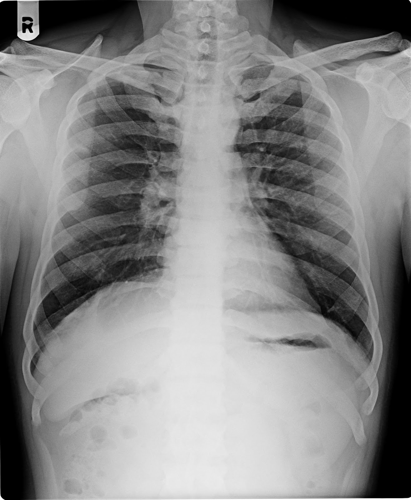
X-RAY CHEST PA VIEW.
Both lung fields appears normal.
No evidence of koch’s lesion or consolidation seen.
Both apices, cardiophrenic, costophrenic angles and domes of the diaphragm are normal.
The cardiac size is within normal limit.
Trachea is central, no mediastinal shift is seen and the mediastinal outlines do not show any
abnormality.
Bony thorax appear normal
IMPRESSION ; NORMAL
X RAY CHEST .
Thanks for reference
DR.BHAVESH PATEL DR.NIRAV
DESAI DR.DEEPAK SHARMA DR.JIGNESH PATEL
MD, DMRE. MB,DMRD MD(Radio diagnosis) MB,DMRD
---------------------------------------------------------------------------------------------------------------------