Saturday 23 September 2017

23/09/17 SALU PANDIT F/25Y CXR-PA


23/09/17 SALU PANDIT F/25Y CXR-PA
TAV KHASHI  LAST 15DAYS

 NAME       :  SALU  PANDIT
AGE        :  25  YRS
REF DR      ; 
DATE        : 23/09/2017

X-RAY CHEST PA VIEW.
? koch's lesion is seen at left upper zone .
Both apices, cardiophrenic, costophrenic angles and domes of  the diaphragm are normal.
The cardiac size is within normal limit.
Trachea is central, no mediastinal shift is seen and the  mediastinal outlines do not show any abnormality.
Bony thorax appear normal

  
Thanks for reference

DR.BHAVESH PATEL     DR.NIRAV DESAI    DR.DEEPAK SHARMA    DR.JIGNESH PATEL
                 MD, DMRE.           MB,DMRD                   MD(Radio diagnosis)                MB,DMRD


------------------------------------------------------------------------------------------------------------
      

Friday 22 September 2017

23/09/17 DRASHTI THAKOR F/2Y CXR-AP

23/09/17 DRASHTI THAKOR F/2Y CXR-AP

H/O CHEKUP
NAME     :    DRASHTI  THAKOR
AGE       :   2   YRS
REF DR     ;   ASTHA  HOSPITAL
DDATE      :   23/09/2017

X-RAY CHEST PA VIEW.

Both lung fields appears normal.
No evidence of koch’s lesion or consolidation seen.
Both apices, cardiophrenic, costophrenic angles and domes of  the diaphragm are normal.
The cardiac size is within normal limit.
Trachea is central, no mediastinal shift is seen and the  mediastinal outlines do not show any abnormality.
Bony thorax appear normal


IMPRESSION ;   NORMAL X RAY CHEST .


Thanks for reference

DR.BHAVESH PATEL     DR.NIRAV DESAI    DR.DEEPAK SHARMA    DR.JIGNESH PATEL
                 MD, DMRE.           MB,DMRD                   MD(Radio diagnosis)                MB,DMRD


------------------------------------------------------------------------------------------------------------

Thursday 21 September 2017

21/09/17 PRATHAM RABARI M/11 MONTH RT.SKULL-LAT

21/09/17 PRATHAM RABARI M/11 MONTH RT.SKULL-LAT

H/O HEAD  INJURY LAST 2 DAYS

NAME   :   PRATHAM
AGE      :  11  MONTHS
REF BY: DR.
 DATE   : 021.09.2017


                                  X-RAY SKULL  LAT. VIEW


The bone density is normal.
No evidence of linear or depressed fracture is seen in skull vault.
No evidence of erosion or sclerosis is seen in skull vault.
Sella turcica appear normal.
No evidence of changes of raised intra cranial tension is seen.


Thanks for reference.                                          
                                                                                               

DR.BHAVESH PATEL                                                   
                 MD, DMRE.                                                                 


------------------------------------------------------------------------------------------------------

Wednesday 20 September 2017

20/09/17 KANTABEN ZALA F/48Y CXR-PA

20/09/17 KANTABEN ZALA F/48Y CXR-PA

H/O TAV  KHASHI  LAST 8 DAYS

-------------- 
NAME   :    KANTABEN
AGE      :    48   Years.
REF BY:     DR.
 DATE   :    20/09/2017


X-RAY CHEST PA VIEW.


Both lung fields shows prominent bronchovascular markings suggest changes of Bronchitis.

No evidence of koch’s lesion or consolidation seen.

Both apices, cardiophrenic, costophrenic angles and domes of  the diaphragm are normal.

The cardiac size is within normal limit.

Trachea is central, no mediastinal shift is seen and the  mediastinal outlines do not show any abnormality.

Bony thorax and soft tissues of the chest wall are normal.


IMPRESSION : - Both lung fields shows prominent bronchovascular markings suggest changes of Bronchitis.


Thanks for reference


DR.BHAVESH PATEL     DR.NIRAV DESAI    DR.DEEPAK SHARMA    DR.JIGNESH PATEL
                 MD, DMRE.           MB,DMRD                   MD(Radio diagnosis)                MB,DMRD

 ----------------------------------------------------------------------------------------------

Tuesday 19 September 2017

19/09/17 RAKESHBHAI CHRISTAIN M/42Y CXR-PA

19/09/17 RAKESHBHAI CHRISTAIN M/42Y CXR-PA

H/O KHASHI LAST 8 DAYS
NAME     :    RAKESHBHAI
AGE        :  42  YRS
REF DR  ;   DR.
DATE      :  19.09.2017

X-RAY CHEST PA VIEW.

Both lung fields appears normal.
No evidence of koch’s lesion or consolidation seen.
Both apices, cardiophrenic, costophrenic angles and domes of  the diaphragm are normal.
The cardiac size is within normal limit.
Trachea is central, no mediastinal shift is seen and the  mediastinal outlines do not show any abnormality.
Bony thorax appear normal


IMPRESSION ;   NORMAL X RAY CHEST .

Thanks for reference

DR.BHAVESH PATEL     DR.NIRAV DESAI    DR.DEEPAK SHARMA    DR.JIGNESH PATEL
                 MD, DMRE.           MB,DMRD                   MD(Radio diagnosis)                MB,DMRD


------------------------------------------------------------------------------------------------------------

Monday 18 September 2017

18/09/17 NANDABEN CHUNARA F/50Y CXR-PA

18/09/17 NANDABEN CHUNARA F/50Y  CXR-PA
H/O PRE OP


NAME       : NANDABEN   CHUNARA
AGE        :50  YRS
REF DR      ;   
DATE      :  18/09/2017

X-RAY CHEST PA VIEW.

Both lung fields appears normal.
No evidence of koch’s lesion or consolidation seen.
Both apices, cardiophrenic, costophrenic angles and domes of  the diaphragm are normal.
The cardiac size is within normal limit.
Trachea is central, no mediastinal shift is seen and the  mediastinal outlines do not show any abnormality.
Bony thorax appear normal


IMPRESSION ;   NORMAL X RAY CHEST .

Thanks for reference

DR.BHAVESH PATEL     DR.NIRAV DESAI    DR.DEEPAK SHARMA    DR.JIGNESH PATEL

                 MD, DMRE.           MB,DMRD                   MD(Radio diagnosis)                MB,DMRD
------------------------------------------------------------------------------------------------------------

Saturday 16 September 2017

16/09/17 SURESHBHAI MARVADI 30Y. ABDOMIN SUPAIN


16/09/17 SURESHBHAI MARVADI 30Y. ABDOMIN SUPAIN

H/O ABDOMIN  API GASTRIK PAIN  LAST 2 DAYS.

NAME      : SURESHBHAI  
AGE     :  30  YEARS
REF BY   :     
DATE     :  16/09/2017

 X-RAY OF ABDOMEN (KUB)
-----------------------------------------------

There is no radio-opaque calculus seen in the region of both kidneys,both ureters and bladder.
No evidence of abnormal dilatation of bowel loops.
Both psoas shadows appear normal.
Visible  Lumbo sacral spine appears normal.


DR.BHAVESH PATEL     DR.NIRAV DESAI    DR.DEEPAK SHARMA    DR.JIGNESH PATEL
                 MD, DMRE.           MB,DMRD                   MD(Radiodiagnosis)                MB,DMRD


------------------------------------------------------------------------------------------------------------

Friday 15 September 2017

16/09/17 MOHINIBEN RATHI F/52Y RT.KNEE-AP/LAT

16/09/17 MOHINIBEN RATHI F/52Y RT.KNEE-AP/LAT

RT.KNEE PAIN LAST 5 DAYS
NAME     :    MOHINIBEN
AGE        :    52   Yrs.
REF BY  : DR.
DATE     : 16 /09/2017


 X-RAY RT KNEE JOINT AP & LAT.


The bone density is normal.

No lytic or sclerotic densities seen.

Rt knee joint space and alignment is maintained.

No evidence of fracture or dislocation seen.

The articular and subarticular margins are smooth with no abnormality seen.

No subarticular abnormality is seen.

The alignment of joint is maintained.

The patellofemoral articulation is normal.

No soft tissue abnormality is seen.


IMPRESSION- Normal Study.

Thanks for reference


DR. JIGNESH  PATEL                                                DR. DEEPAK  SHARMA
             MB, DMRD                                                         MD ( Radiodiagnosis).


------------------------------------------------------------------------------------------------------------

Thursday 14 September 2017

14/09/17 URVISH MAHESHWARI M/22Y CXR-PA

14/09/17 URVISH MAHESHWARI M/22Y CXR-PA

H/O LT.SIDE LOWAR CHEST PAIN LAST 3 DAYS
NAME         :     URVISH  MAHESHWARI
AGE        :      22   YRS
REF DR      ;      DR.
DATE        :       14.09.2017

X-RAY CHEST PA VIEW.

Both lung fields appears normal.
No evidence of koch’s lesion or consolidation seen.
Both apices, cardiophrenic, costophrenic angles and domes of  the diaphragm are normal.
The cardiac size is within normal limit.
Trachea is central, no mediastinal shift is seen and the  mediastinal outlines do not show any abnormality.
Bony thorax appear normal


IMPRESSION ;   NORMAL X RAY CHEST .

Thanks for reference

DR.BHAVESH PATEL     DR.NIRAV DESAI    DR.DEEPAK SHARMA    DR.JIGNESH PATEL
                 MD, DMRE.           MB,DMRD                   MD(Radio diagnosis)                MB,DMRD


------------------------------------------------------------------------------------------------------------

Wednesday 13 September 2017

13/09/17 AJAY RAVAL M/17 Y CXR-PA

13/09/17 AJAY RAVAL M/17 Y CXR-PA

H/O  SWASH/KHASHI/TAV  LAST 8 DAYS
NAME     :   AJAY RAVAL
AGE        :  17   YRS
REF DR  ;   DR.
DATE      :   13.09.2017

X-RAY CHEST PA VIEW.

Both lung fields appears normal.
No evidence of koch’s lesion or consolidation seen.
Both apices, cardiophrenic, costophrenic angles and domes of  the diaphragm are normal.
The cardiac size is within normal limit.
Trachea is central, no mediastinal shift is seen and the  mediastinal outlines do not show any abnormality.
Bony thorax appear normal


IMPRESSION ;   NORMAL X RAY CHEST .

Thanks for reference

DR.BHAVESH PATEL     DR.NIRAV DESAI    DR.DEEPAK SHARMA    DR.JIGNESH PATEL
                 MD, DMRE.           MB,DMRD                   MD(Radio diagnosis)                MB,DMRD


------------------------------------------------------------------------------------------------------------
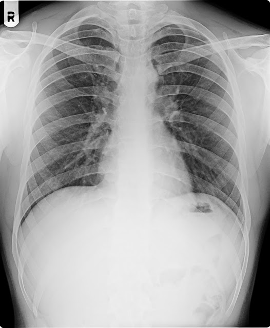
13/09/17 MANJULABEN BARAIYA F/28Y CXR-PA

13/09/17 MANJULABEN BARAIYA F/28Y CXR-PA

H/O LT.SIDE CHEST PAIN LAST 4 DAYS
NAME     :    MANJULABEN
AGE        :  28   YRS
REF DR  ;   DR.
DATE      :   13.09.2017

X-RAY CHEST PA VIEW.

Both lung fields appears normal.
No evidence of koch’s lesion or consolidation seen.
Both apices, cardiophrenic, costophrenic angles and domes of  the diaphragm are normal.
The cardiac size is within normal limit.
Trachea is central, no mediastinal shift is seen and the  mediastinal outlines do not show any abnormality.
Bony thorax appear normal


IMPRESSION ;   NORMAL X RAY CHEST .

Thanks for reference

DR.BHAVESH PATEL     DR.NIRAV DESAI    DR.DEEPAK SHARMA    DR.JIGNESH PATEL
                 MD, DMRE.           MB,DMRD                   MD(Radio diagnosis)                MB,DMRD


------------------------------------------------------------------------------------------------------------

Monday 11 September 2017

11/09/17 BHAIJIBHAI BHOI M/50Y CXR-PA

11/09/17 BHAIJIBHAI BHOI M/50Y CXR-PA

H/O FEVAR  KHASHI  SWASH LAST 5 DAYS


X-RAY CHEST PA VIEW.

Both lung fields shows prominent bronchovascular markings suggest changes of Bronchitis.

No evidence of koch’s lesion or consolidation seen.

Both apices, cardiophrenic, costophrenic angles and domes of  the diaphragm are normal.

The cardiac size is within normal limit.

Trachea is central, no mediastinal shift is seen and the  mediastinal outlines do not show any abnormality.

Bony thorax and soft tissues of the chest wall are normal.


IMPRESSION : - Both lung fields shows prominent bronchovascular markings suggest changes of Bronchitis.


Thanks for reference

DR.BHAVESH PATEL     DR.NIRAV DESAI    DR.DEEPAK SHARMA    DR.JIGNESH PATEL
                 MD, DMRE.           MB,DMRD                   MD(Radio diagnosis)                MB,DMRD


------------------------------------------------------------------------------------------------------------

11/09/17 BHOMIK TADVI M/11 MONTH RT.HAND-AP

11/09/17 BHOMIK TADVI M/11 MONTH RT.HAND-AP

H/O ENJQRY LAST 3 DAYS

NAME:    BHOMIK
AGE    : 11 MONTH
DATE :   11/09/2017
X-RAY RIGHT HAND AP & OBLIQUE


Bone density is normal.

No evidence of fracture or dislocation seen.

No lytic/sclerotic lesion is seen.

The distal ends of radius, ulna and visualised carpal bones shows a normal anatomic
configuration and alignment.

The joint spaces are maintained & no obvious articular or  subarticular lesion is seen.

Soft tissue planes appear swelling.


Thanks for reference


DR.BHAVESH PATEL                                                DR. DEEPAK  SHARMA
             MD, DMRE.                                                         MD ( Radiodiagnosis).


------------------------------------------------------------------------------------------------------------

Sunday 10 September 2017

11/09/17 AMITKUMAR DABHI M/25Y CXR-PA

11/09/17 AMITKUMAR DABHI M/25Y CXR-PA

H/O RUTAIN CHEKUP
NAME   :    amitkumar
AGE      :     25  Years.
REF BY:     DR.
 DATE   :    11.09.2017


X-RAY CHEST PA VIEW.


Both lung fields shows prominent bronchovascular markings suggest changes of Bronchitis.
Azygos vein seen in RT upper lobe.

No evidence of koch’s lesion or consolidation seen.

Both apices, cardiophrenic, costophrenic angles and domes of  the diaphragm are normal.

The cardiac size is within normal limit.

Trachea is central, no mediastinal shift is seen and the  mediastinal outlines do not show any abnormality.

Bony thorax and soft tissues of the chest wall are normal.


IMPRESSION : - Both lung fields shows prominent bronchovascular markings suggest changes of Bronchitis.


Thanks for reference


DR.BHAVESH PATEL     DR.NIRAV DESAI    DR.DEEPAK SHARMA    DR.JIGNESH PATEL
                 MD, DMRE.           MB,DMRD                   MD(Radio diagnosis)                MB,DMRD


------------------------------------------------------------------------------------------------------------

10/09/17 BHIKHABHAI RAVAL M/47Y CXR-PA

10/09/17 BHIKHABHAI RAVAL M/47Y CXR-PA

H/O COUGH/ HAEMOPTYSIS/FEVAR LAST 4 DAYS

------------------------------------------------------------------------------------------------------------

Wednesday 6 September 2017

07/09/17 MAHI BHARGAV F/20 MONTH CXR-AP/B.HIPS-AP


07/09/17 MAHI BHARGAV F/20 MONTH CXR-AP/B.HIPS-AP


H/O NOT ABLE TO WALK


 NAME     :  MAHI
AGE        : 20  MONTHS.
REF DR  ;     
DATE      : 07/09/2017

X-RAY CHEST PA VIEW.

Both lung fields appears normal.
No evidence of koch’s lesion or consolidation seen.
Both apices, cardiophrenic, costophrenic angles and domes of  the diaphragm are normal.
The cardiac size is enlarged .
Trachea is central, no mediastinal shift is seen and the  mediastinal outlines do not show any abnormality.
Bony thorax appear normal


X-RAY OF PELVIS  WITH BOTH HIP JOINTS  -AP VIEW


-The bone density is normal.

-Visible bony pelvis appear normal.

-No e/o hip joint dislocation is seen.no fracture is seen .

-No evidence of erosion or sclerosis is seen.

-Articular margins and joint space appear normal.
  
Thanks for reference

DR.BHAVESH PATEL     DR.NIRAV DESAI    DR.DEEPAK SHARMA    DR.JIGNESH PATEL
                 MD, DMRE.           MB,DMRD                   MD(Radio diagnosis)                MB,DMRD


------------------------------------------------------------------------------------------------------------

06/09/17 POOJABEN PATEL F/36Y CT.SPINE-SPI-AP/LAT


06/09/17 POOJBEN PATEL F/36Y CERVICO THORA-SPI-AP/LAT

H/O LT SIDE SHOULDER PAIN LAST 1 MONTH

NAME   :   POOJBEN
AGE       :  36   Years
REF BY : DR.
DATE    :   06/09/2017

X-RAY CERVICAL SPINE AP & LATERAL VIEW

The bone density is normal.

No obvious lytic or sclerotic lesion.

The  cervical  vertebral bodies and their  appendages  (pedicles and spinous  process) are normal.

The intervertebral disc spaces are maintained with normal heights and
no subchondral bony changes.

The intervertebral joints do not show any abnormality.

The cranio vertebral junction is normal.

The  spinal alignment is maintained (with normal  lordotic  curve and
alignment of the anterior/posterior spinal lines).

Pre and para vertebral soft tissues do not show any abnormality.

No evidence of bony cervical ribs seen on either side.


IMPRESSION   :   NORMAL STUDY  


 X-RAY DORSO-LUMBER SPINE  LATERAL VIEW

The bone density is normal.

No obvious lytic or sclerotic lesion.

Minimal degenerative changes in form of osteophytes seen in dorsal spine.

The  dorso-lumber  vertebral bodies and their  appendages  (pedicles, spinous and
transverse process) are normal.

The intervertebral disc spaces are maintained with normal heights and
no subchondral bony changes.

No evidence of lysis or lysthesis seen.

The intervertebral joints do not show any abnormality.

The  spinal alignment is maintained.

Pre and paravertebral soft tissues do not show any abnormality.


Thanks for reference


DR.BHAVESH PATEL     DR.NIRAV DESAI    DR.DEEPAK SHARMA    DR.JIGNESH PATEL
                 MD, DMRE.           MB,DMRD                   MD(Radio diagnosis)                MB,DMRD


------------------------------------------------------------------------------------------------------------

Tuesday 5 September 2017

05/09/17 LEELABEN THAKOR F/43Y CXR-PA

05/09/17  LEELABEN THAKOR F/43Y CXR-PA

H/O FEVAR  COUGHFING LAST 15 DAYS
NAME   :    LEELABEN  THAKOR
AGE      :    43  YEARS.
REF BY:     DR.
 DATE   :    05/09/2017 


X-RAY CHEST PA VIEW.


Homogenous opacity seen in RT upper and mid zone suggest consolidation possibility of Kochs aetiology.

Both cardiophrenic, costophrenic angles and domes of  the diaphragm are normal.

The cardiac size is within normal limit.

Trachea is central, no mediastinal shift is seen and the  mediastinal outlines do not show any abnormality.

Bony thorax and soft tissues of the chest wall are normal.


IMPRESSION : - Homogenous opacity seen in RT upper and mid zone suggest consolidation possibility of Kochs aetiology.


Thanks for reference


DR.BHAVESH PATEL     DR.NIRAV DESAI    DR.DEEPAK SHARMA    DR.JIGNESH PATEL
                 MD, DMRE.           MB,DMRD                   MD(Radio diagnosis)                MB,DMRD


------------------------------------------------------------------------------------------------------------

05/09/17 RIMA S. MAITY F/26Y CXR-PA

05/09/17 RIMA S. MAITY F/26Y CXR-PA

H/O RUTIAN CHEK UP
NAME     :    RIMA  S. MAITY
AGE        :  26  YRS
REF DR  ;   DR.
DATE      :   05/09/2017

X-RAY CHEST PA VIEW.

Both lung fields appears normal.
No evidence of koch’s lesion or consolidation seen.
Both apices, cardiophrenic, costophrenic angles and domes of  the diaphragm are normal.
The cardiac size is within normal limit.
Trachea is central, no mediastinal shift is seen and the  mediastinal outlines do not show any abnormality.
Bony thorax appear normal


IMPRESSION ;   NORMAL X RAY CHEST .

Thanks for reference

DR.BHAVESH PATEL     DR.NIRAV DESAI    DR.DEEPAK SHARMA    DR.JIGNESH PATEL
                 MD, DMRE.           MB,DMRD                   MD(Radio diagnosis)                MB,DMRD


------------------------------------------------------------------------------------------------------------