Sunday 31 December 2017

01/01/18 DAHIBEN PATEL F/75Y CERVICAL-AP/LAT


01/01/18 DAHIBEN PATEL F/75Y CERVICAL-AP/LAT
H/O NECK PAIN LAST ONE MONTH


NAME   : DAHIBEN  PATEL
AGE       :75  Years
REF BY : DR.
DATE    : 01/01/2018

X-RAY CERVICAL SPINE AP & LATERAL VIEW

The bone density is normal.

No obvious lytic or sclerotic lesion.

The  cervical  vertebral bodies and their  appendages  (pedicles and spinous  process) are normal.

The intervertebral disc spaces are maintained with normal heights and
no subchondral bony changes.

The intervertebral joints do not show any abnormality.

The cranio vertebral junction is normal.

The  spinal alignment is maintained (with normal  lordotic  curve and
alignment of the anterior/posterior spinal lines).

Pre and para vertebral soft tissues do not show any abnormality.

No evidence of bony cervical ribs seen on either side.


IMPRESSION:NORMAL STUDY


Thanks for reference

DR.BHAVESH PATEL     DR.NIRAV DESAI    DR.DEEPAK SHARMA    DR.JIGNESH PATEL
                 MD, DMRE.           MB,DMRD                   MD(Radio diagnosis)                MB,DMRD


--------------------------------------------------------------------------------------------------------------------------

Friday 29 December 2017

30/12/17 KOKILABEN PATEL F/60Y CXR-PA

30/12/17 KOKILABEN PATEL F/60Y CXR-PA

H/O KHASHI  FEVAR  LAST 20 DAYS
NAME     :    KOKILABEN  PATEL
AGE        :  60  YRS
REF DR  ;   DR.
DATE      :   30.12.2017

X-RAY CHEST PA VIEW.

Both lung fields appears normal.
No evidence of koch’s lesion or consolidation seen.
Both apices, cardiophrenic, costophrenic angles and domes of  the diaphragm are normal.
The cardiac size is within normal limit.
Trachea is central, no mediastinal shift is seen and the  mediastinal outlines do not show any abnormality.
Bony thorax appear normal


IMPRESSION ;   NORMAL X RAY CHEST .

Thanks for reference

DR.BHAVESH PATEL     DR.NIRAV DESAI    DR.DEEPAK SHARMA    DR.JIGNESH PATEL
                 MD, DMRE.           MB,DMRD                   MD(Radio diagnosis)                MB,DMRD


--------------------------------------------------------------------------------------------------------------------------

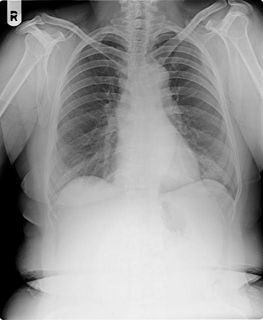
30/12/17 BABUBHAI SOLANKI M/40Y CXR-PA/LT.CXR-LAT


30/12/17 BABUBHAI SOLANKI M/40Y CXR-PA/LT.CXR-LAT
H/O TRUMA LAST 2 DAYS LT.SIDELOWAR CHEST PAIN

NAME     :     BABUBHAI                                  
AGE        :  40   YRS
REF DR  ;   DR.
DATE   :  30.12.2017

X-RAY CHEST PA VIEW.

Both lung fields appears normal.
No evidence of koch’s lesion or consolidation seen.
Both apices, cardiophrenic, costophrenic angles and domes of  the diaphragm are normal.
The cardiac size is within normal limit.
Trachea is central, no mediastinal shift is seen and the  mediastinal outlines do not show any abnormality.
Bony thorax appear normal


IMPRESSION ;   NORMAL X RAY CHEST .

Thanks for reference

DR.BHAVESH PATEL     DR.NIRAV DESAI    DR.DEEPAK SHARMA    DR.JIGNESH PATEL
                 MD, DMRE.           MB,DMRD                   MD(Radio diagnosis)                MB,DMRD


--------------------------------------------------------------------------------------------------------------------------


Wednesday 27 December 2017

27/12/17 M.SHAMIM ANSHARI M/57Y CXR-PA

27/12/17 M.SHAMIM ANSHARI M/57Y CXR-PA

H/O KHASHI LAST 3 MONTH
NAME   :   M. SHAMIN 
AGE      :   57    Years.
REF BY:     DR.
 DATE   :   27.12.2017 


X-RAY CHEST PA VIEW.


Both lung fields shows prominent bronchovascular markings suggest changes of Bronchitis.

No evidence of koch’s lesion or consolidation seen.

Both apices, cardiophrenic, costophrenic angles and domes of  the diaphragm are normal.

The cardiac size is within normal limit.

Trachea is central, no mediastinal shift is seen and the  mediastinal outlines do not show any abnormality.

Bony thorax and soft tissues of the chest wall are normal.


IMPRESSION : - Both lung fields shows prominent bronchovascular markings suggest changes of Bronchitis.


Thanks for reference


DR.BHAVESH PATEL     DR.NIRAV DESAI    DR.DEEPAK SHARMA    DR.JIGNESH PATEL
                 MD, DMRE.           MB,DMRD                   MD(Radio diagnosis)                MB,DMRD


------------------------------------------------------------------------------------------------------------

Tuesday 26 December 2017

27/12/17 HEMSINH RATHOD M/55Y CXR-PA/CERVICAL-AP/LAT


27/12/17 HEMSINH RATHOD M/55Y CXR-PA/CERVICAL-AP/LAT
H/O TRUMA PAIN LT.NECK&LT.U.CHEST 6 DAYS


NAME    : HEMSINH  RATHOD
AGE       : 55  Years
REF BY : .
DATE    :  27/12/2017

X-RAY CERVICAL SPINE AP & LATERAL VIEW

The bone density is normal.

No obvious lytic or sclerotic lesion.

The  cervical  vertebral bodies and their  appendages  (pedicles and spinous  process) are normal.

The intervertebral disc spaces are maintained with normal heights and
no subchondral bony changes.

The intervertebral joints do not show any abnormality.

The cranio vertebral junction is normal.

The  spinal alignment is maintained (with normal  lordotic  curve and
alignment of the anterior/posterior spinal lines).

Pre and para vertebral soft tissues do not show any abnormality.

No evidence of bony cervical ribs seen on either side.


IMPRESSION:NORMAL STUDY


X-RAY CHEST PA VIEW.

Both lung fields appears normal.

No evidence of koch’s lesion or consolidation seen.

Both apices, cardiophrenic, costophrenic angles and domes of  the diaphragm are normal.

The cardiac size is within normal limit.

Trachea is central, no mediastinal shift is seen and the  mediastinal outlines do not show any abnormality.

Bony thorax appears normal.


IMPRESSION : - Normal Study.

Thanks for reference


DR.BHAVESH PATEL     DR.NIRAV DESAI    DR.DEEPAK SHARMA    DR.JIGNESH PATEL
                 MD, DMRE.           MB,DMRD                   MD(Radio diagnosis)                MB,DMRD


------------------------------------------------------------------------------------------------------------

Friday 22 December 2017

22/12/17 BHARTIBEN THAKOR F/32Y LT.KNEE-AP/LAT

22/12/17 BHARTIBEN THAKOR F/32Y LT.KNEE-AP/LAT
H/O LT.KNEE PAIN LAST 1YRS
  
NAME   : BHARTIBEN  THAKOR
AGE      : 32  YEARS.
REF BY  : 
DATE     : 22/12/2017
                                       
                                     
 X-RAY LT KNEE JOINT AP & LAT.


The bone density is normal.

No lytic or sclerotic densities seen.

Lt knee joint space and alignment is maintained.

The articular and subarticular margins are smooth with no abnormality seen.

No subarticular abnormality is seen.

The alignment of joint is maintained.

The patellofemoral articulation is normal.

No soft tissue abnormality is seen.


IMPRESSION-NORMAL STUDY .


Thanks for reference


DR.JIGNESH  PATEL                                           
            MB, DMRD.                                         


------------------------------------------------------------------------------------------------------------

22/12/17 PRABHATBHAI RAVAD M/34Y CXR-PA

22/12/17 PRABHATBHAI RAVAD M/34Y CXR-PA
H/O KHASHI LAST 7 DAYS

NAME   :    PRABHATBHAI
AGE      :    34   Years.
REF BY:     
 DATE   :    22/12/2017

X-RAY CHEST PA VIEW.


Both lung fields shows prominent bronchovascular markings suggest changes of Bronchitis.

No evidence of koch’s lesion or consolidation seen.

Both apices, cardiophrenic, costophrenic angles and domes of  the diaphragm are normal.

The cardiac size is within normal limit.

Trachea is central, no mediastinal shift is seen and the  mediastinal outlines do not show any abnormality.

Bony thorax and soft tissues of the chest wall are normal.


IMPRESSION : - Both lung fields shows prominent bronchovascular markings suggest changes of Bronchitis.


Thanks for reference


DR.BHAVESH PATEL     DR.NIRAV DESAI    DR.DEEPAK SHARMA    DR.JIGNESH PATEL
                 MD, DMRE.           MB,DMRD                   MD(Radio diagnosis)                MB,DMRD


------------------------------------------------------------------------------------------------------------

Wednesday 20 December 2017

21/12/17 HARDIK PATEL M/21Y LT.MENDIBLE

21/12/17 HARDIK PATEL M/21Y LT.MENDIBLE

H/O LT.SIDE MENDIBLE PAIN LAST 10 DAYS

NAME   :    HARDIK PATEL
AGE       :    21 YEARS
REF DR ;     
DATE     :   21.12.2017

X RAY OF  LT MENDIBLE  ;

No fracture or dislocation seen.
Bones & joints under view appear normal.
No evidence of  osteolytic  or osteosclerotic lesion is seen .
No evidence of  abnormal vascular calcification seen.


DR.BHAVESH PATEL     DR.NIRAV DESAI    DR.DEEPAK SHARMA    DR.JIGNESH PATEL
                 MD, DMRE.           MB,DMRD                   MD(Radio diagnosis)                MB,DMRD


------------------------------------------------------------------------------------------------------------

20/12/17 PALLAVI SENGAR F/6Y CXR-PA

20/12/17 PALLAVI SENGAR F/6Y CXR-PA

H/O KHASHI LAST TWO MONTH

NAME    :    PALLAVI  SENGAR
AGE       : 6   YRS
REF DR  ;   DR.
DATE     :   20/12/2017

X-RAY CHEST PA VIEW.

Both lung fields appears normal.
No evidence of koch’s lesion or consolidation seen.
Both apices, cardiophrenic, costophrenic angles and domes of  the diaphragm are normal.
The cardiac size is within normal limit.
Trachea is central, no mediastinal shift is seen and the  mediastinal outlines do not show any abnormality.
Bony thorax appear normal


IMPRESSION ;   NORMAL X RAY CHEST .

Thanks for reference

DR.BHAVESH PATEL     DR.NIRAV DESAI    DR.DEEPAK SHARMA    DR.JIGNESH PATEL
                 MD, DMRE.           MB,DMRD                   MD(Radio diagnosis)                MB,DMRD


------------------------------------------------------------------------------------------------------------

Tuesday 19 December 2017

19/12/17 ZEEL SUTHAR M/19Y CXRPA

19/12/17  ZEEL SUTHAR M/19Y CXRPA

H/O TAV  KHASHI LAST ONE MONTH

NAME     :ZEEL  SUTHAR
AGE        : 22  YRS
REF DR  ;   DR.
DATE      :  19/12/2017

X-RAY CHEST PA VIEW.

Both lung fields appears normal.
No evidence of koch’s lesion or consolidation seen.
Both apices, cardiophrenic, costophrenic angles and domes of  the diaphragm are normal.
The cardiac size is within normal limit.
Trachea is central, no mediastinal shift is seen and the  mediastinal outlines do not show any abnormality.
Bony thorax appear normal


IMPRESSION ;   NORMAL X RAY CHEST .

Thanks for reference

DR.BHAVESH PATEL     DR.NIRAV DESAI    DR.DEEPAK SHARMA    DR.JIGNESH PATEL
                 MD, DMRE.           MB,DMRD                   MD(Radio diagnosis)                MB,DMRD


------------------------------------------------------------------------------------------------------------

Sunday 17 December 2017

18/12/17 JIVABHAI THAKOR M/60Y CXR-PA

18/12/17 JIVABHAI THAKOR M/60Y CXR-PA

TAV  KHASHI  LAST  15 DAYS

NAME            :    JIVABHAI  THAKOR
AGE               :    60   Years.
REF BY         :     DR.
 DATE            :   18/12/2017


X-RAY CHEST PA VIEW.


Fibrocalcified kochs lesion seen in RT upper zone.
Homogenous opacity seen in RT upper zone with shift of trachea towards RT side suggest RT Upper lobe collapse.
Calcified kochs lesion seen in LT mid zone.
Minimal bronchiectatic changes seen in LT lower zone.


Both  cardiophrenic, costophrenic angles and domes of  the diaphragm are normal.

The cardiac size is within normal limit.


Bony thorax and soft tissues of the chest wall are normal.


IMPRESSION : - Fibrocalcified kochs lesion seen in RT upper zone.
Homogenous opacity seen in RT upper zone with shift of trachea towards RT side suggest RT Upper lobe collapse.
Calcified kochs lesion seen in LT mid zone.
Minimal bronchiectatic changes seen in LT lower zone.


Thanks for reference


DR.BHAVESH PATEL     DR.NIRAV DESAI    DR.DEEPAK SHARMA    DR.JIGNESH PATEL
                 MD, DMRE.           MB,DMRD                   MD(Radio diagnosis)                MB,DMRD


------------------------------------------------------------------------------------------------------------

17/12/17 RAVI CHAUHAN M/20Y CXR-PA

17/12/17 RAVI CHAUHAN M/20Y CXR-PA
H/O RT.SIDE CHEST PAIN LAST 15 DAYS

NAME            :   RAVI  CHAUHAN  
AGE               :   20 YEARS.
REF BY         :   DR.
 DATE            :  17/12/2017


X-RAY CHEST PA VIEW.


Kochs infiltration seen in  Both upper zone.

Homogenous opacity seen in RT mid and lower zone obscuring RT costophrenic angle suggest RT pleural effusion.

LT costophrenic angle is  minimally blunted possibility of Minimal LT pleural effusion.


The cardiac size is within normal limit.

Trachea is central, no mediastinal shift is seen and the  mediastinal outlines do not show any abnormality.

Bony thorax and soft tissues of the chest wall are normal.


IMPRESSION : - Kochs infiltration seen in  Both upper zone.
-Homogenous opacity seen in RT mid and lower zone obscuring RT costophrenic angle suggest RT pleural effusion.
-LT costophrenic angle is  minimally blunted possibility of Minimal LT pleural effusion.


Thanks for reference


DR.BHAVESH PATEL     DR.NIRAV DESAI    DR.DEEPAK SHARMA    DR.JIGNESH PATEL
                 MD, DMRE.           MB,DMRD                   MD(Radio diagnosis)                MB,DMRD


------------------------------------------------------------------------------------------------------------

17/12/17 MAKANBHAI SARVAIYA M/65Y CXR-PA

17/12/17 MAKANBHAI SARVAIYA M/65Y CXR-PA
H/O TAV  KHASHI  LAST ONE MONTH


NAME            :    MAKANBHAI
AGE               :    65   Years.
REF BY         :     
 DATE            :     17/12/2017

                                                       X-RAY CHEST PA VIEW.


Both lung fields shows prominent bronchovascular markings suggest changes of Bronchitis.

No evidence of koch’s lesion or consolidation seen.

Both apices, cardiophrenic, costophrenic angles and domes of  the diaphragm are normal.

The cardiac size is within normal limit.

Trachea is central, no mediastinal shift is seen and the  mediastinal outlines do not show any abnormality.

Bony thorax and soft tissues of the chest wall are normal.


IMPRESSION : - Both lung fields shows prominent bronchovascular markings suggest changes of Bronchitis.

  
Thanks for reference
  
DR.BHAVESH PATEL     DR.NIRAV DESAI    DR.DEEPAK SHARMA    DR.JIGNESH PATEL
                 MD, DMRE.           MB,DMRD                   MD(Radio diagnosis)                MB,DMRD


------------------------------------------------------------------------------------------------------------

Friday 15 December 2017

15/12/17 SHAILESH ZALA M/25Y CXR-PA

15/12/17 SHAILESH ZALA M/25Y CXR-PA
MOUTH BLEDING LAST 4 DAYS


NAME   :    SHAILESHBHAI  ZALA
AGE      :    25  Years.
REF BY:    
 DATE   :   15/12/2017
X-RAY CHEST PA VIEW.


Both lung fields shows prominent bronchovascular markings suggest changes of Bronchitis.

No evidence of koch’s lesion or consolidation seen.

Both apices, cardiophrenic, costophrenic angles and domes of  the diaphragm are normal.

The cardiac size is within normal limit.

Trachea is central, no mediastinal shift is seen and the  mediastinal outlines do not show any abnormality.

Bony thorax and soft tissues of the chest wall are normal.


IMPRESSION : - Both lung fields shows prominent bronchovascular markings suggest changes of Bronchitis.


Thanks for reference


DR.BHAVESH PATEL     DR.NIRAV DESAI    DR.DEEPAK SHARMA    DR.JIGNESH PATEL
                 MD, DMRE.           MB,DMRD                   MD(Radio diagnosis)                MB,DMRD


------------------------------------------------------------------------------------------------------------

Wednesday 13 December 2017

13/12/17 MUKESHBHAI PARMAR M/31Y CERVICAL SPI-AP

13/12/17 MUKESHBHAI PARMAR M/31Y CERVICAL SPI-AP

H/O PAIN IN THROOT
NAME   :  MUKESHBHAI
AGE       :  31  Years
REF BY : DR.
DATE    :  13.12.2017

X-RAY CERVICAL SPINE AP

The bone density is normal.

No obvious lytic or sclerotic lesion.

The  cervical  vertebral bodies and their  appendages  (pedicles and spinous  process) are normal.

The intervertebral disc spaces are maintained with normal heights and
no subchondral bony changes.

The intervertebral joints do not show any abnormality.

The cranio vertebral junction is normal.

The  spinal alignment is maintained (with normal  lordotic  curve and
alignment of the anterior/posterior spinal lines).

para vertebral soft tissues do not show any abnormality.

No evidence of bony cervical ribs seen on either side.


IMPRESSION  :   NORMAL STUDY

Thanks for reference

DR.BHAVESH PATEL     DR.NIRAV DESAI    DR.DEEPAK SHARMA    DR.JIGNESH PATEL
                 MD, DMRE.           MB,DMRD                   MD(Radio diagnosis)                MB,DMRD


------------------------------------------------------------------------------------------------------------

Tuesday 12 December 2017

12/12/17 SAURABH TIWARI M/24Y CXR-PA

12/12/17 SAURABH TIWARI M/24Y CXR-PA

H/O SWASH  KHASHI  LAST 4 MONTH

NAME   :    SAURABH
AGE      :    24    Years.
REF BY:     DR.
 DATE   :   12.12.2017


X-RAY CHEST PA VIEW.


Both lung fields shows prominent bronchovascular markings suggest changes of Bronchitis.

No evidence of koch’s lesion or consolidation seen.

Both apices, cardiophrenic, costophrenic angles and domes of  the diaphragm are normal.

The cardiac size is within normal limit.

Trachea is central, no mediastinal shift is seen and the  mediastinal outlines do not show any abnormality.

Bony thorax and soft tissues of the chest wall are normal.


IMPRESSION : - Both lung fields shows prominent bronchovascular markings suggest changes of Bronchitis.


Thanks for reference


DR.BHAVESH PATEL     DR.NIRAV DESAI    DR.DEEPAK SHARMA    DR.JIGNESH PATEL
                 MD, DMRE.           MB,DMRD                   MD(Radio diagnosis)                MB,DMRD


------------------------------------------------------------------------------------------------------------

12/12/17 SHRI LOCHAN BHOI M/30Y CXR-PA

12/12/17 SHRI LOCHAN BHOI M/30Y CXR-PA

H/O TAV  KHASHI  SWASH LAST 6 DAYS

NAME   :    SHRI LOCHAN BHOI
AGE      :     30   Years.
REF BY:     DR.
 DATE   :     12/12/2017


X-RAY CHEST PA VIEW.


Both lung fields shows prominent bronchovascular markings suggest changes of Bronchitis.

No evidence of koch’s lesion or consolidation seen.

Both apices, cardiophrenic, costophrenic angles and domes of  the diaphragm are normal.

The cardiac size is within normal limit.

Trachea is central, no mediastinal shift is seen and the  mediastinal outlines do not show any abnormality.

Bony thorax and soft tissues of the chest wall are normal.


IMPRESSION : - Both lung fields shows prominent bronchovascular markings suggest changes of Bronchitis.


Thanks for reference


DR.BHAVESH PATEL     DR.NIRAV DESAI    DR.DEEPAK SHARMA    DR.JIGNESH PATEL
                 MD, DMRE.           MB,DMRD                   MD(Radio diagnosis)                MB,DMRD


------------------------------------------------------------------------------------------------------------

Sunday 10 December 2017

11/12/17 HARDEV GUJJAR M/6Y CXR-PA

11/12/17 HARDEV GUJJAR M/6Y CXR-PA

H/O SWASH LAST 5YRS
NAME     :    HARDEV  GAJJAR
AGE        : 6   YRS
REF DR  ;   DR.
DATE      :   11.12.2017

X-RAY CHEST PA VIEW.

Bilateral hilum prominence .
No evidence of koch’s lesion or consolidation seen.
Both apices, cardiophrenic, costophrenic angles and domes of  the diaphragm are normal.
The cardiac size is within normal limit.
Trachea is central, no mediastinal shift is seen and the  mediastinal outlines do not show any abnormality.
Bony thorax appear normal


IMPRESSION ;   
 Bilateral hilum prominence .

Thanks for reference

DR.BHAVESH PATEL     DR.NIRAV DESAI    DR.DEEPAK SHARMA    DR.JIGNESH PATEL
                 MD, DMRE.           MB,DMRD                   MD(Radio diagnosis)                MB,DMRD


------------------------------------------------------------------------------------------------------------

Friday 8 December 2017

09/12/17 HANSABEN RATHOD F/26Y RT.HAND

09/12/17 HANSABEN RATHOD F/26Y RT.HAND
H/O RT.HAND SWALING

NAME    :   KAILASHBEN PATEL                       
AGE       :    50 Years/F
REF BY  : SHREE SAI MEDICAL CENTRE
DATE     :  09/12/2017

X-RAY RIGHT HAND AP & OBLIQUE


Bone density is normal.

No evidence of fracture or dislocation seen.

No lytic/sclerotic lesion is seen.

The distal ends of radius, ulna and visualised carpal bones shows a normal anatomic
configuration and alignment.

The joint spaces are maintained & no obvious articular or  subarticular lesion is seen.

Soft tissue planes appear normal.


Thanks for reference


 DR. DEEPAK  SHARMA
       MD ( Radio diagnosis).


------------------------------------------------------------------------------------------------------------

Sunday 3 December 2017

04/12/17 NISHITH PATEL M/25Y CXR-PA

04/12/17 NISHITH PATEL M/25Y CXR-PA

H/O COMPANY FITNESH
NAME     :   NISHITH
AGE        :  25   YRS
REF DR  ;   DR.
DATE      :   04.12.20177

X-RAY CHEST PA VIEW.

Both lung fields appears normal.
No evidence of koch’s lesion or consolidation seen.
Both apices, cardiophrenic, costophrenic angles and domes of  the diaphragm are normal.
The cardiac size is within normal limit.
Trachea is central, no mediastinal shift is seen and the  mediastinal outlines do not show any abnormality.
Bony thorax appear normal


IMPRESSION ;   NORMAL X RAY CHEST .

Thanks for reference

DR.BHAVESH PATEL     DR.NIRAV DESAI    DR.DEEPAK SHARMA    DR.JIGNESH PATEL
                 MD, DMRE.           MB,DMRD                   MD(Radio diagnosis)                MB,DMRD

--------------------------------------------------------------------------------------------------------------------------