Wednesday 28 February 2018

01\03\18 KHODABHAI THAKOR M\52Y CXR-PA


01\03\18 KHODABHAI THAKOR M\52Y CXR-PA
H\O OLD COX , THREE DAYS MOUTH BLIDING.


NAME   :    khodabhai   thakor    
AGE       :   52   Years
REF DR ;     
DATE     :  01.03.2018


X-RAY OF CHEST (P.A VIEW)                                                
------------------------------------------------

Koch’s infiltration is seen at both upper and mid zones .

Rest of the lung fields appear normal.
Both costo-phrenic angles appear normal.
No evidence of hilar or mediastinal lymph adenopathy.
Heart size appears normal.
Bony thoracic cage appears normal.


DR.JIGNESH PATEL
MB DMRD

--------------------------------------------------------------------------------------------------------------------

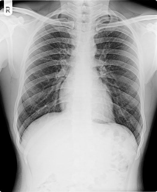
01\03\18 AKHILESH KUMAR M\23Y CXR-PA

01\03\18 AKHILESH KUMAR M\23Y CXR-PA
H\O KHASHI LAST 8 DAYS

NAME     :   AKHILESH KUMAR
AGE        :  23  YRS
REF DR  ;     DR.
DATE      :   01.03.2018

X-RAY CHEST PA VIEW.

Both lung fields appears normal.
No evidence of koch’s lesion or consolidation seen.
Both apices, cardiophrenic, costophrenic angles and domes of  the diaphragm are normal.
The cardiac size is within normal limit.
Trachea is central, no mediastinal shift is seen and the  mediastinal outlines do not show any abnormality.
Bony thorax appear normal


IMPRESSION ;   NORMAL X RAY CHEST .

Thanks for reference

DR.BHAVESH PATEL     DR.NIRAV DESAI    DR.DEEPAK SHARMA    DR.JIGNESH PATEL
                 MD, DMRE.           MB,DMRD                   MD(Radio diagnosis)                MB,DMRD


--------------------------------------------------------------------------------------------------------------------------

Thursday 22 February 2018

22/02/18 YOGANSH PARMAR M/13Y CXR-PA

22/02/18 YOGANSH  PARMAR M/13Y CXR-PA
H/O KHASHI LAST 8 DAYS


NAME   :    YOGANSH
AGE      :     13 Years.
REF BY:     DR.
 DATE   :    22.02.2018


X-RAY CHEST PA VIEW.


Both lung fields shows prominent bronchovascular markings suggest changes of Bronchitis.

No evidence of koch’s lesion or consolidation seen.

Both apices, cardiophrenic, costophrenic angles and domes of  the diaphragm are normal.

The cardiac size is within normal limit.

Trachea is central, no mediastinal shift is seen and the  mediastinal outlines do not show any abnormality.

Bony thorax and soft tissues of the chest wall are normal.


IMPRESSION : - Both lung fields shows prominent bronchovascular markings suggest changes of Bronchitis.


Thanks for reference


DR.BHAVESH PATEL     DR.NIRAV DESAI    DR.DEEPAK SHARMA    DR.JIGNESH PATEL
                 MD, DMRE.           MB,DMRD                   MD(Radio diagnosis)                MB,DMRD


--------------------------------------------------------------------------------------------------------------------------

Wednesday 21 February 2018

21/2/18 JASVANTBHAI DANTANI 33Y, CXR.

21/2/18 JASVANTBHAI DANTANI 33Y, CXR.

H/O ONE WWEEK FEVAR,CHEST PAIN,COUGHING.


NAME   :    jaswantbhai
AGE      :    33  Years.
REF BY:     
 DATE   :    21.02.2018


X-RAY CHEST PA VIEW.


Both lung fields shows prominent bronchovascular markings suggest changes of Bronchitis.

No evidence of koch’s lesion or consolidation seen.

Both apices, cardiophrenic, costophrenic angles and domes of  the diaphragm are normal.

The cardiac size is within normal limit.

Trachea is central, no mediastinal shift is seen and the  mediastinal outlines do not show any abnormality.

Bony thorax and soft tissues of the chest wall are normal.


IMPRESSION : - Both lung fields shows prominent bronchovascular markings suggest changes of Bronchitis.


DR.BHAVESH PATEL     DR.NIRAV DESAI    DR.DEEPAK SHARMA    DR.JIGNESH PATEL
                 MD, DMRE.           MB,DMRD                   MD(Radio diagnosis)                MB,DMRD

------------------------------------------------------------------------------------------------------------------------

21/2/18 ANIS AHMED 34M CXR

21/2/18 ANIS AHMED 34M CXR
H/O ONE MONTH COUGHING, PAST HISTRY COX.


NAME   :    ANIS AHEMAD
AGE      :    34   Years.
REF BY:     DR.
 DATE   :    21.02.2018


X-RAY CHEST PA VIEW.


Fibrocalcified kochs lesion seen in Both Upper and Mid zone.
Both lung fields shows prominent bronchovascular markings suggest changes of Bronchitis.

No evidence of active koch’s lesion or consolidation seen.

Both apices, cardiophrenic, costophrenic angles and domes of  the diaphragm are normal.

The cardiac size is within normal limit.

Trachea is central, no mediastinal shift is seen and the  mediastinal outlines do not show any abnormality.

Bony thorax and soft tissues of the chest wall are normal.


IMPRESSION : -Fibrocalcified kochs lesion seen in Both Upper and Mid zone.
Both lung fields shows prominent bronchovascular markings suggest changes of Bronchitis.


Thanks for reference


DR.BHAVESH PATEL     DR.NIRAV DESAI    DR.DEEPAK SHARMA    DR.JIGNESH PATEL
                 MD, DMRE.           MB,DMRD                   MD(Radio diagnosis)                MB,DMRD


--------------------------------------------------------------------------------------------------------------------------

Thursday 15 February 2018

16/02/18 RAMESHBHAI RATHOD M/27Y CXR-PA

16/02/18 RAMESHBHAI RATHOD M/27Y CXR-PA
H/O FEVAR  KHASHI  LAST ONE MONTH


NAME   :    RAMESHBHAI  RATHOD
AGE      :    27  Years.
REF BY:     
 DATE   :    16/02/2018


X-RAY CHEST PA VIEW.


Both lung fields shows prominent bronchovascular markings suggest changes of Bronchitis.

No evidence of koch’s lesion or consolidation seen.

Both apices, cardiophrenic, costophrenic angles and domes of  the diaphragm are normal.

The cardiac size is within normal limit.

Trachea is central, no mediastinal shift is seen and the  mediastinal outlines do not show any abnormality.

Bony thorax and soft tissues of the chest wall are normal.


IMPRESSION : - Both lung fields shows prominent bronchovascular markings suggest changes of Bronchitis.


Thanks for reference


DR.BHAVESH PATEL     DR.NIRAV DESAI    DR.DEEPAK SHARMA    DR.JIGNESH PATEL
                 MD, DMRE.           MB,DMRD                   MD(Radio diagnosis)                MB,DMRD


--------------------------------------------------------------------------------------------------------------------------

Tuesday 13 February 2018

13/02/18 MADANLAL MANGALIYA M/35Y CXR-PA

13/02/18 MADANLAL MANGALIYA M/35Y CXR-PA

H/O FEVAR  COUGH  CHEST PAIN  LAST 5 DAYS
NAME   :    MADANLAL  MANGALIYA
AGE      :    35    Years.
REF BY:     DR.
 DATE   :     13.02.2018


X-RAY CHEST PA VIEW.


Consolidation / ?? Kochs seen in LT lower zone .

The cardiac size is within normal limit.

Trachea is central, no mediastinal shift is seen and the  mediastinal outlines do not show any abnormality.

Bony thorax and soft tissues of the chest wall are normal.


Thanks for reference


DR.BHAVESH PATEL     DR.NIRAV DESAI    DR.DEEPAK SHARMA    DR.JIGNESH PATEL
                 MD, DMRE.           MB,DMRD                   MD(Radio diagnosis)                MB,DMRD

Sunday 11 February 2018

11/02/18 ALPESH THAKOR M/19Y CXR-PA

11/02/18 ALPESH THAKOR M/19Y CXR-PA
H/O FEVAR  COUGHFING  CHEST PAIN  LAST 15 DAYS

NAME   :    ALPESH
AGE      :    19    Years.
REF BY:     DR.
 DATE   :    11/02/2018


X-RAY CHEST PA VIEW.


Kochs infiltration seen in RT upper and mid zone and in RT perihilar region and in LT mid zone in LT perihilar region.

The cardiac size is within normal limit.

Trachea is central, no mediastinal shift is seen and the  mediastinal outlines do not show any abnormality.

Bony thorax and soft tissues of the chest wall are normal.


IMPRESSION : - Kochs infiltration seen in RT upper and mid zone and in RT perihilar region and in LT mid zone in LT perihilar region.


Thanks for reference


DR.BHAVESH PATEL     DR.NIRAV DESAI    DR.DEEPAK SHARMA    DR.JIGNESH PATEL
                 MD, DMRE.           MB,DMRD                   MD(Radio diagnosis)                MB,DMRD


--------------------------------------------------------------------------------------------------------------------------

Saturday 10 February 2018

11/02/18 KAJAL RAJPUT F/15Y CXR-PA

11/02/18 KAJAL RAJPUT F/15Y CXR-PA
H/O FEVAR LAST 2 DAYS


NAME   :    KAJAL  RAJPUT
AGE      :   15 Years.
REF BY:   
 DATE   :   11/02/201


X-RAY CHEST PA VIEW.


Both lung fields shows prominent bronchovascular markings 

No evidence of koch’s lesion or consolidation seen.

Both apices, cardiophrenic, costophrenic angles and domes of  the diaphragm are normal.

The cardiac size is within normal limit.

Trachea is central, no mediastinal shift is seen and the  mediastinal outlines do not show any abnormality.

Bony thorax and soft tissues of the chest wall are normal.


IMPRESSION : - Both lung fields shows prominent bronchovascular markings 


Thanks for reference


DR.BHAVESH PATEL     DR.NIRAV DESAI    DR.DEEPAK SHARMA    DR.JIGNESH PATEL
                 MD, DMRE.           MB,DMRD                   MD(Radio diagnosis)                MB,DMRD


--------------------------------------------------------------------------------------------------------------------------

Thursday 8 February 2018

09/02/18 SANTOSH KUMAR M/30Y CXR-PA

09/02/18 SANTOSH KUMAR M/30Y CXR-PA
H/O KHASHI  TAV LAST 8 DAYS

NAME   :   SANTOSH KUMAR  
AGE      :    30 Years.
REF BY: 
    DATE   :  09/02/2018

                                    X-RAY CHEST PA VIEW.


Both lung fields shows prominent bronchovascular markings suggest changes of Bronchitis.

No evidence of koch’s lesion or consolidation seen.

Both apices, cardiophrenic, costophrenic angles and domes of  the diaphragm are normal.

The cardiac size is within normal limit.

Trachea is central, no mediastinal shift is seen and the  mediastinal outlines do not show any abnormality.

Bony thorax and soft tissues of the chest wall are normal.


IMPRESSION : - Both lung fields shows prominent bronchovascular markings suggest changes of Bronchitis.


Thanks for reference


DR.BHAVESH PATEL     DR.NIRAV DESAI    DR.DEEPAK SHARMA    DR.JIGNESH PATEL
                 MD, DMRE.           MB,DMRD                   MD(Radio diagnosis)                MB,DMRD


--------------------------------------------------------------------------------------------------------------------------

Wednesday 7 February 2018

07/02/18 RAJUBEN OOD F/35Y CXR-PA

07/02/18 RAJUBEN OOD F/35Y CXR-PA

H/O CHEST PAIN LAST 3 DAYS
NAME    :  RAJUBEN OOD
AGE   :  35  YEARS
DR. :
DATE   :  07.02.2018

X-RAY CHEST PA VIEW.


Bronchiectatic changes with surrounding consolidation are noted in right paracardiac region.

Both apices, costophrenic angles and domes of  the diaphragm are normal.

The cardiac size is within normal limit.

Trachea is central, no mediastinal shift is seen and the  mediastinal outlines do not show any abnormality.

Bony thorax and soft tissues of the chest wall are normal.


Thanks for reference

DR.BHAVESH PATEL     DR.NIRAV DESAI    DR.DEEPAK SHARMA    DR.JIGNESH PATEL
                 MD, DMRE.           MB,DMRD                   MD(Radio diagnosis)         

--------------------------------------------------------------------------------------------------------------------------

Tuesday 6 February 2018

06/02/18 JAYDEEP BHOI M/25Y CXR-PA

06/02/18 JAYDEEP BHOI M/25Y CXR-PA

H/O KHASHI LAST 20 DAYS

-------------------------------------------------------------------------------------------------------------------------- 

NAME   : JAYDEEP         
AGE       :       Years
REF DR ;   
DATE     :  06.02.2018


X-RAY OF CHEST (P.A VIEW)                                                
------------------------------------------------

Fibro calcified old koch's lesion is seen in RT upper zone.
RT c p angle is blunted suggest pleural thickening or ?? effusion.

Rest of the lung fields appear normal.
LT costo-phrenic angle appear normal.
No evidence of hilar or mediastinal lymph adenopathy.
Heart size appears normal.
Bony thoracic cage appears normal.


Monday 5 February 2018

05/02/15 PUNIT YADAV M/24Y CXR-PA

05/02/15 PUNIT YADAV M/24Y CXR-PA

H/O KHASHI LAST 10 DAYS

X-RAY CHEST PA VIEW.


Both lung fields appears normal.
No evidence of koch’s lesion or consolidation seen.

Both apices, cardiophrenic, costophrenic angles and domes of  the diaphragm are normal.

The cardiac size is within normal limit.

Trachea is central, no mediastinal shift is seen and the  mediastinal outlines do not show any abnormality.

Bony thorax and soft tissues of the chest wall are normal.


IMPRESSION : - Normal Study.

Thanks for reference


DR.BHAVESH PATEL     DR.NIRAV DESAI    DR.DEEPAK SHARMA    DR.JIGNESH PATEL

                 MD, DMRE.           MB,DMRD                   MD(Radio diagnosis)                MB,DMRD
--------------------------------------------------------------------------------------------------------------------------